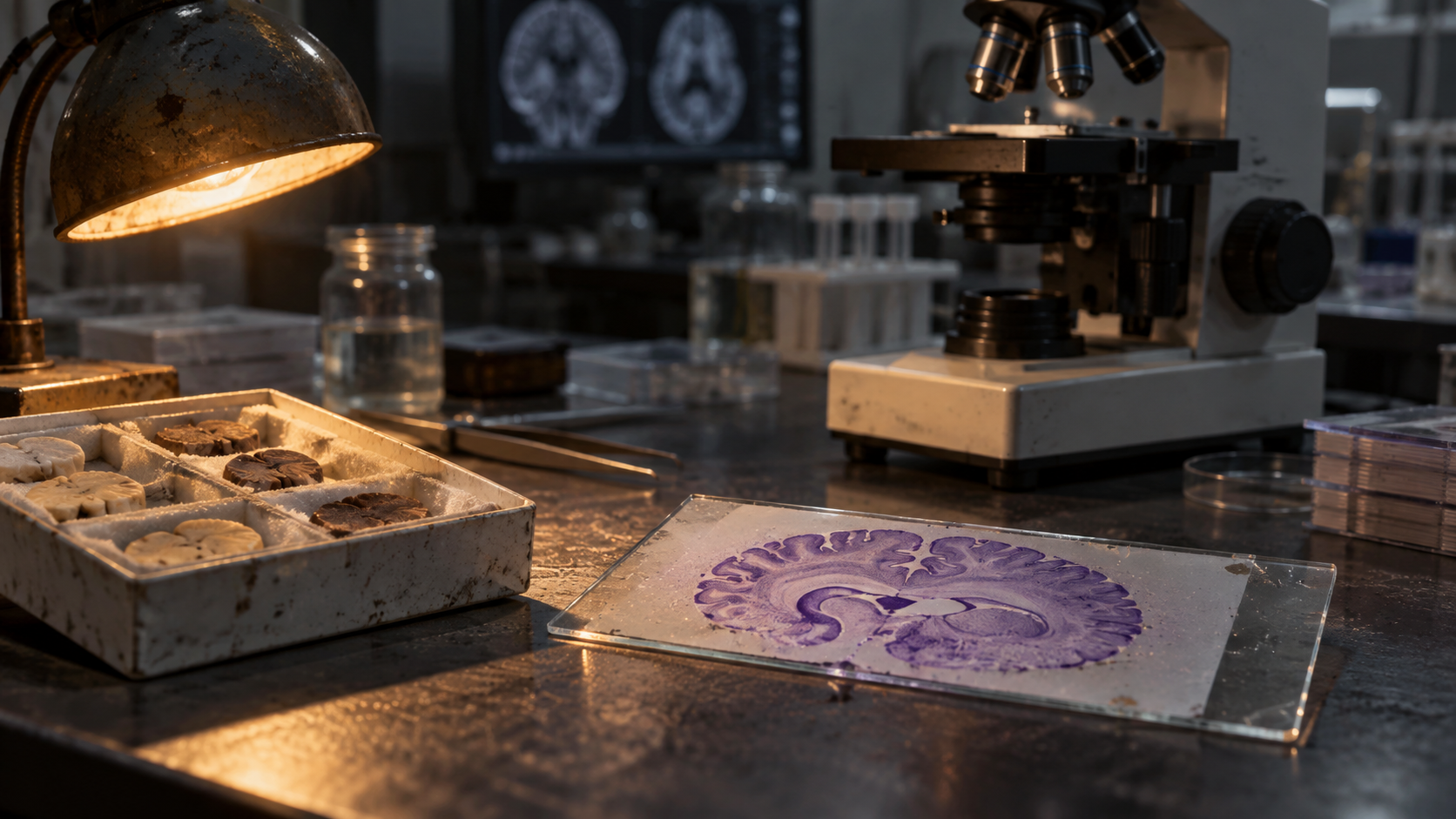
Inside the atlas
A research team analyzed brain tissue from 1,494 donors to create high-resolution molecular maps of Alzheimer’s disease and related brain disorders. The dataset includes 6.3 million cell nuclei from the prefrontal cortex, a region involved in memory, planning and other cognitive functions.
By examining gene activity at the level of individual cell nuclei, researchers mapped how different cell types respond across disease groups and stages. The atlas can help scientists compare molecular patterns that may be obscured when tissue is analyzed as a whole.
What the maps can and cannot do
The resource is intended to guide basic and translational research. It may help identify cell populations and pathways for further study, but it does not establish that any one molecular pattern causes disease or points to an effective treatment.
The atlas is not a clinical diagnostic test. It does not predict an individual’s future health, and the findings do not provide a new therapy. Additional studies are needed to test candidate mechanisms in independent samples and determine whether they can inform treatment research.
Sources
NIH-sourced molecular brain atlas (2026) built from 1,494 donors and 6.3 million prefrontal-cortex cell nuclei, as stated in the cited NIH report.
The source is cited only as a general 'NIH, Molecular Brain Atlas Report, 2026' with no DOI or direct link to a peer-reviewed paper.
The full publication name/DOI is not given; the exact list of 'related disorders' covered beyond Alzheimer's is not specified in the article; response from the contacted institution not yet received.